| Colic flexures | |
|---|---|
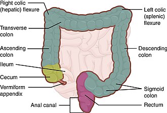 The hepatic and splenic flexures labelled at either side of transverse colon | |
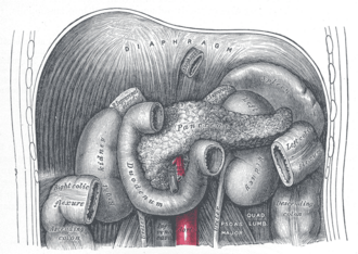 | |
| Details | |
| Precursor | Hindgut |
| Artery | Right colic artery (right flexure), and left colic artery (left flexure) |
| Identifiers | |
| Latin | flexura coli |
| FMA | 14555 |
| Anatomical terminology | |
In the anatomy of the human digestive tract, there are two colic flexures, or curvatures in the transverse colon. The right colic flexure is also known as the hepatic flexure, and the left colic flexure is also known as the splenic flexure.1 There are colic flexures between adjacent segments of the colon, which can interfere with the course of a colonoscopy.2 Some authors consider it necessary to clarify and recognize the terminology related to "colic flexures."3
Structure
Right colic flexure
The right colic flexure or hepatic flexure (as it is next to the liver) is the sharp bend between the ascending colon and the transverse colon. The hepatic flexure lies in the right upper quadrant of the human abdomen. It receives blood supply from the superior mesenteric artery.
Left colic flexure
The left colic flexure or splenic flexure (as it is close to the spleen) is the sharp bend between the transverse colon and the descending colon. The splenic flexure receives dual blood supply from the terminal branches of the superior mesenteric artery and the inferior mesenteric artery.4
Clinical significance
The splenic flexure is the last and highest positioned flexure in the colon. Gas can build up at this flexure and give abdominal pain giving rise to a condition known as splenic flexure syndrome. Splenic flexure syndrome is often found in those with irritable bowel syndrome (IBS), and is considered by some practitioners to be a type of IBS since it can also result from stress.5
The splenic flexure is a watershed region as it receives dual blood supply from the terminal branches of the superior mesenteric artery and the inferior mesenteric artery, thus making it prone to ischemic damage in cases of low blood pressure because it does not have its own primary source of blood. In the context of bowel ischemia in particular ischemic colitis, the splenic flexure is sometimes referred to as Griffith's point, along with the upper rectum (Sudeck's point).46
Additional images
-
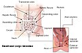 Intestines
 -
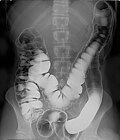 Double contrast barium enema, using positive and negative contrast
References
References
- Jones, Jeremy (17 March 2009). "Transverse colon". Radiopaedia. Retrieved 2 January 2022.
- Wozniak, Slawomir; Pawlus, Aleksander; Grzelak, Joanna; Chobotow, Slawomir; Paulsen, Friedrich; Olchowy, Cyprian; Zaleska-Dorobisz, Urszula (January 2023). "Acute colonic flexures: the basis for developing an artificial intelligence-based tool for predicting the course of colonoscopy". Anatomical Science International. 98 (1): 136–142. doi:10.1007/s12565-022-00681-8. ISSN 1447-6959.
- Wozniak, Slawomir; Quondamatteo, Fabio; Paulsen, Friedrich (April 2023). "Flexures and bends of the large intestine: Current terminology and a suggestion to simplify it". Journal of Anatomy. 242 (4): 695–700. doi:10.1111/joa.13800. ISSN 0021-8782.
- Dixon, Andrew. "Griffiths point". Radiopaedia. Retrieved 2 January 2022.
- "What Is the Splenic Flexure?". WebMD. Retrieved 2 January 2022.
- Amini, Afshin; Nagalli, Shivaraj (31 July 2023). "Bowel Ischemia". StatPearls Publishing. PMID 32119414. Retrieved 17 October 2025.
External links
External links
- Lotti M. Anatomy in relation to left colectomy
- Anatomy photo:37:13-0102 at the SUNY Downstate Medical Center
- Anatomy image:8182 at the SUNY Downstate Medical Center