A scanning helium ion microscope (SHIM, HeIM or HIM) is a high-resolution imaging instrument that uses a scanning helium ion beam to visualize at sub-nanometer resolution. It was developed in the mid-2000s for nanotechnology applications, and has been demonstrated to have a better surface resolution than scanning electron microscopes.
Description
Scanning helium ion microscope (SHIM) was developed in the mid-2000s as a new microscopy technique for nanotechnology applications.2 It uses a focused beam of helium ions to scan a specimen's surface instead of electrons used in a scanning electron microscope (SEM).34 It utilizes a gas field ionization source (GFIS), which is used to produce a highly focused beam of helium ions.5
The very high source brightness, and the short De Broglie wavelength of the helium ions, enables extremely small probe sizes.45 Similar to other focused ion beam techniques, it allows to combine milling and cutting of samples with their observation at sub-nanometer resolution.56 A surface resolution of 0.24 nanometers has been demonstrated by Zeiss.78
When the helium ions interact with a sample under visualization, it does not suffer from large excitation volume, and hence produces a much smaller interaction volume compared to electrons, leading to better surface sensitivity. These characteristics enable the SHIM to obtain qualitative data with a greater resolution that is not achievable with conventional microscopes which use photons or electrons as the emitting source.4 The small interaction volume results in high contrast images, with a large depth of field on a wide range of materials.2 The microscope is also better at imaging insulating materials because it causes less charging on interaction with such materials compared to electron beams.4
History
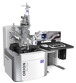
The SHIMs were made commercially available by Zeiss in 2007, and the first microscope was shipped to the National Institute of Standards and Technology.9 However, it was discontinued by Zeiss in 2023.10
References
References
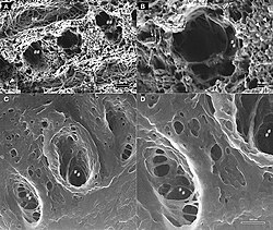
- Bidlack, Felicitas B.; Huynh, Chuong; Marshman, Jeffrey; Goetze, Bernhard (2014). "Helium ion microscopy of enamel crystallites and extracellular tooth enamel matrix". Frontiers in Physiology. 5: 395. doi:10.3389/fphys.2014.00395. PMC 4193210. PMID 25346697.
- Postek, Michael T.; Vladar, Andras; Ming, Bin (March 2009). "Understanding Imaging and Metrology with the Helium Ion Microscope". National Institute of Standards and Technology. pp. 249–260. Retrieved June 17, 2025.
- "ALIS Corporation Announces Breakthrough in Helium Ion Technology for Next-Generation Atomic-Level Microscope". December 7, 2005. Archived from the original on May 28, 2006. Retrieved November 22, 2008.
- Postek, Michael T.; Vladar, Andras; Kramar, John A.; Stern, L. A.; Notte, John; McVey, Sean (January 2007). "Helium Ion Microscopy: A New Technique for Semiconductor Metrology and Nanotechnology". National Institute of Standards and Technology. pp. 161–167. Retrieved June 17, 2025.
- "Helium ion microscope offers unprecedented resolution". Oak Ridge National Laboratory. Retrieved June 17, 2025.
- Iberi, Vighter; Vlassiouk, Ivan; Zhang, X.-G.; Matola, Brad; Linn, Allison; Joy, David C.; Rondinone, Adam J. (2015). "Maskless Lithography and in situ Visualization of Conductivity of Graphene using Helium Ion Microscopy". Scientific Reports. 5 11952. Bibcode:2015NatSR...511952I. doi:10.1038/srep11952. PMC 4493665. PMID 26150202.
- "Microscopy resolution record claimed by Carl Zeiss". Fabtech.org. November 21, 2008. Archived from the original on January 8, 2009. Retrieved November 22, 2008.
- "Carl Zeiss Sets New World Record in Microscopy Resolution Using Scanning Helium Ions" (Press release). November 21, 2008. Archived from the original on May 1, 2009. Retrieved November 22, 2008.
- "Carl Zeiss SMT Ships World's First ORION Helium Ion Microscope to U.S. National Institute of Standards and Technology". Zeiss (Press release). July 17, 2008. Retrieved November 22, 2008.
{{cite press release}}: CS1 maint: deprecated archival service (link) - "Discontinued ZEISS Microscopy Products". Zeiss. Retrieved April 25, 2025.
External links
External links
- Carl Zeiss SMT – Nano Technology Systems Division: ORION He-Ion microscope
- Microscopy Today, Volume 14, Number 04, July 2006: An Introduction to the Helium Ion Microscope
- How New Helium Ion Microscope Measures Up – ScienceDaily