Article · Wikipedia archive·Last revised Jul 20, 2026
Tricarboxylate transport protein, mitochondrial
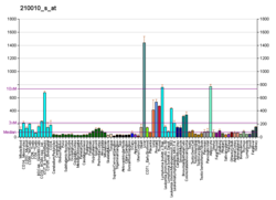
Tricarboxylate transport protein, mitochondrial, also known as tricarboxylate carrier protein and citrate transport protein (CTP), is a protein that in humans is encoded by the SLC25A1 gene. SLC25A1 belongs to the mitochondrial carrier gene family SLC25. High levels of the tricarboxylate transport protein are found in the liver, pancreas and kidney. Lower or no levels are present in the brain, heart, skeletal muscle, placenta and lung.
Tricarboxylate transport protein, mitochondrial, also known as tricarboxylate carrier protein and citrate transport protein (CTP), is a protein that in humans is encoded by the SLC25A1gene.5678 SLC25A1 belongs to the mitochondrial carrier gene family SLC25.91011 High levels of the tricarboxylate transport protein are found in the liver, pancreas and kidney. Lower or no levels are present in the brain, heart, skeletal muscle, placenta and lung.911
The tricarboxylate transport protein is located within the inner mitochondria membrane. It provides a link between the mitochondrial matrix and cytosol by transporting citrate through the impermeable inner mitochondrial membrane in exchange for malate from the cytosol.9101112 The citrate transported out of the mitochondrial matrix by the tricarboxylate transport protein is catalyzed by citrate lyase to acetyl CoA, the starting material for fatty acid biosynthesis, and oxaloacetate.10 As well, cytosolic NADPH + H+ necessary for fatty acid biosynthesis is generated in the reduction of oxaloacetate to malate and pyruvate by malate dehydrogenase and the malic enzyme.111314 For these reasons, the tricarboxylate transport protein is considered to play a key role in fatty acid synthesis.10
Structure
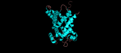
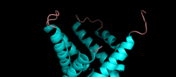
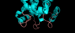
A 3D cartoon depiction of the tripartite structure of a mitochondrial transport protein, generated from 23CE bovine mitochondrial ADP-ATP carriersource ↗A zoomed in image of the C and N termini and the two loops linking the repeated domains on the cytoplasmic side of the inner mitochondrial membrane. source ↗A zoomed in image of the three loops linking the two α-helices of each repeated domain located on the matrix side of the membrane. source ↗
The structure of the tricarboxylate transport protein is consistent with the structures of other mitochondrial carriers.91012 In particular, the tricarboxylate transport protein has a tripartite structure consisting of three repeated domains that are approximately 100 amino acids in length.912 Each repeat forms a transmembrane domain consisting of two hydrophobic α-helices.91015 The amino and carboxy termini are located on the cytosolic side of the inner mitochondrial membrane.910 Each domain is linked by two hydrophilic loops located on the cytosolic side of the membrane.9101516 The two α-helices of each repeated domain are connected by hydrophilic loops located on the matrix side of the membrane.91016 A salt bridge network is present on both the matrix side and cytoplasmic side of the tricarboxylate transport protein.16
Transport mechanism
The tricarboxylate transport protein exists in two states: a cytoplasmic state where it accepts malate from the cytoplasm and a matrix state where it accepts citrate from the mitochondrial matrix.17 A single binding site is present near the center of the cavity of the tricarboxylate transport protein, which can be either exposed to the cytosol or the mitochondrial matrix depending on the state.151617 A substrate induced conformational change occurs when citrate enters from the matrix side and binds to the central cavity of the tricarboxylate transport protein.9 This conformational change opens a gate on the cytosolic side and closes the gate on the matrix side.9 Likewise, when malate enters from the cytosolic side, the matrix gate opens and the cytosolic gate closes.9 Each side of the transporter is open and closed by the disruption and formation of the salt bridge networks, which allows access to the single binding site.1516171819
Disease relevance
Mutations in this gene have been associated with the inborn error of metabolism combined D-2- and L-2-hydroxyglutaric aciduria,20 which was the first reported case of a pathogenic mutation of the SLC25A1 gene.1621 Patients with D-2/L-2-hydroxyglutaric aciduria display neonatal onset metabolic encephalopathy, infantile epilepsy, global developmental delay, muscular hypotonia and early death.162122 It is believed low levels of citrate in the cytosol and high levels of citrate in the mitochondria caused by the impaired citrate transport plays a role in the disease.1622 In addition, increased expression of the tricarboxylate transport protein has been linked to cancer112324 and the production of inflammatory mediators.252627 Therefore, it has been suggested that inhibition of the tricarboxylate transport protein may have a therapeutic effect in chronic inflammation diseases and cancer.26
"Human PubMed Reference:". National Center for Biotechnology Information, U.S. National Library of Medicine.
"Mouse PubMed Reference:". National Center for Biotechnology Information, U.S. National Library of Medicine.
Heisterkamp N, Mulder MP, Langeveld A, ten Hoeve J, Wang Z, Roe BA, et al. (September 1995). "Localization of the human mitochondrial citrate transporter protein gene to chromosome 22Q11 in the DiGeorge syndrome critical region". Genomics. 29 (2): 451–456. doi:10.1006/geno.1995.9982. PMID8666394.
Iacobazzi V, Lauria G, Palmieri F (September 1997). "Organization and sequence of the human gene for the mitochondrial citrate transport protein". DNA Sequence. 7 (3–4): 127–139. doi:10.3109/10425179709034029. PMID9254007.
Palmieri F (April 2013). "The mitochondrial transporter family SLC25: identification, properties and physiopathology". Molecular Aspects of Medicine. 34 (2–3): 465–484. doi:10.1016/j.mam.2012.05.005. PMID23266187.
Palmieri F (February 2004). "The mitochondrial transporter family (SLC25): physiological and pathological implications". Pflügers Archiv: European Journal of Physiology. 447 (5): 689–709. doi:10.1007/s00424-003-1099-7. PMID14598172. S2CID25304722.
Hoffmann GF, Köckler S (2016). "Cerebral Organic Acid Disorders and Other Disorders of Lysine Catabolism". In Saudubray JM, Baumgartner M, Walter J (eds.). Inborn Metabolic Diseases. Germany: Springer. p. 344. ISBN978-3-662-49771-5.
Cohen I, Staretz-Chacham O, Wormser O, Perez Y, Saada A, Kadir R, et al. (February 2018). "A novel homozygous SLC25A1 mutation with impaired mitochondrial complex V: Possible phenotypic expansion". American Journal of Medical Genetics. Part A. 176 (2): 330–336. doi:10.1002/ajmg.a.38574. PMID29226520. S2CID6953669.
Gong W, Emanuel BS, Collins J, Kim DH, Wang Z, Chen F, et al. (June 1996). "A transcription map of the DiGeorge and velo-cardio-facial syndrome minimal critical region on 22q11". Human Molecular Genetics. 5 (6): 789–800. CiteSeerX10.1.1.539.9441. doi:10.1093/hmg/5.6.789. PMID8776594.
Bonofiglio D, Santoro A, Martello E, Vizza D, Rovito D, Cappello AR, et al. (June 2013). "Mechanisms of divergent effects of activated peroxisome proliferator-activated receptor-γ on mitochondrial citrate carrier expression in 3T3-L1 fibroblasts and mature adipocytes". Biochimica et Biophysica Acta (BBA) - Molecular and Cell Biology of Lipids. 1831 (6): 1027–1036. doi:10.1016/j.bbalip.2013.01.014. hdl:11586/65706. PMID23370576.