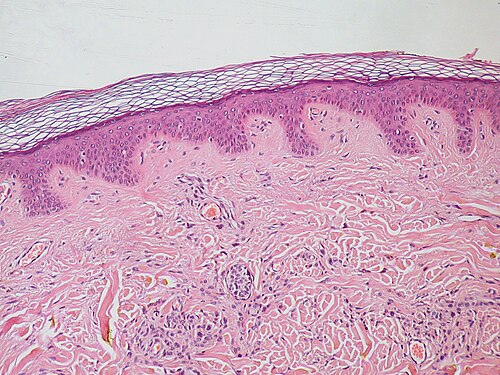
Rete pegs (also known as rete processes, rete ridges or epidermal ridges) are the epithelial extensions that project into the underlying connective tissue in both skin and mucous membranes.
In the epithelium of the mouth, the attached gingiva exhibit rete pegs, while the sulcular2 and junctional epithelia do not.3 Scar tissue lacks rete pegs and scars tend to shear off more easily than normal tissue as a result.1
Also known as papillae, they are downward thickenings of the epidermis between the dermal papillae.
References
References
- Ira D. Papel (2011). Facial Plastic and Reconstructive Surgery (Third ed.). USA: Thieme Medical Publishers. p. 7. ISBN 9781588905154.
- Itoiz, ME; Carranza, FA: The Gingiva. In Newman, MG; Takei, HH; Carranza, FA; editors: Carranza’s Clinical Periodontology, 9th Edition. Philadelphia: W.B. Saunders Company, 2002. pages 23.
- Page, RC; Schroeder, HE. "Pathogenesis of Inflammatory Periodontal Disease: A Summary of Current Work." Lab Invest 1976;34(3):235-249